


Diagnosis and Treatment of Gallstones
Prof JC Sanasam *
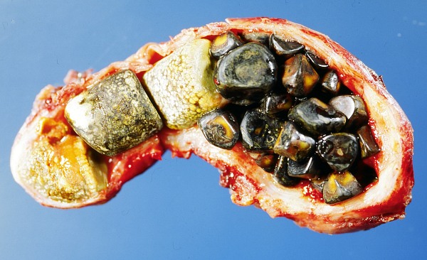
Opened gall bladder containing numerous gallstones :: Pix - wikipedia/Emmanuelm
In the majority of cases gall stones are discovered by accident when the patient is being investigated and treatment begun for something else. Most often a doctor detects it during an ultrasound scan of the abdomen; may suspect of it when he finds cholesterol high in the blood, or even on plain X-ray of the abdomen.
Last week we placed in this column an attempted showcase of what gallstones are, how they are formed and what symptoms they produce in patients who have developed them in their gallbladders. It will not be complete if some highlights on its diagnosis and treatment to be followed are not dealt with.
In the majority of cases gall stones are discovered by accident when the patient is being investigated and treatment begun for something else. Most often a doctor detects it during an ultrasound scan of the abdomen; may suspect of it when he finds cholesterol high in the blood, or even on plain X-ray of the abdomen.
Further investigations in the blood and others can be undertaken in search of signs of evidences that indicate presence of associated or complicated infection, obstruction, pancreatitis, or jaundice.
The following terms may be unintelligible and too technological, but it will be worthwhile just to brush on it so that you will remember when you have to face these procedures and understand the process of the investigations that you may have to undergo.
Cholangiography
In this procedure a dye (which is radio-opaque) is either injected into the blood stream so that it concentrates into the bile ducts and gallbladder, or the dye is inserted straight into the bile ducts through an endoscope (during an ERCP procedure i.e. Endoscopic Retrograde Cholangiopancreatolography) then X-rays are taken to show it on the films. ERCP itself is a procedure to locate as well as to remove stone (as an operational technique).
Normally the dye is supposed to reach the liver, bile ducts, intestines and gall bladder. If the dye is not present in one of these areas it generally means that the gallstone is causing a blockage. Experts can locate also the gallstone single or multiple from this study.
CT scan
This is a study of series of X-rays taken at different axial lines in various planes to locate the gallstones and to know if there is involvement of adjoining tissues and organs.
Cholescintigraphy
A small amount of harmless radioactive material is injected into the patient. This is absorbed by the gallbladder, which is then stimulated to contract. This test can diagnose abnormal contractions of the gallbladder or an obstruction of the bile duct.
Treatment of Gallstones
If symptomless and gallstones are small, no treatment may be needed except taking to restricting oneself to low fat diet.
Cholecystectomy
This is the surgical removal of gallbladder. In earlier days a long incision to open the abdomen wide had to be done. In modern surgery the keyhole surgery (minimally invasive surgery) has become very popular among the surgeons as well as among the patients. If a patient's gallbladder is severely inflamed or it has affected the pancreas it is advisable to opt the open surgery. In advanced centres of many countries a robotic assistant is being used for surgical precision and dexterity in such laparoscopic cholecystectomy.
Ursodeoxycholic acid
Dissolution of gallstones with the medication of this drug can be used for patients who are not suitable for surgery and general anaesthesia. The result takes long up to 24 months. But it is not as effective as surgery.
ERCP
An electrically heated wire is guided through a flexible endoscope inserted through the mouth is used to dilate the opening of the bile duct to remove the stone or to push it down into the intestine.
Lithotripsy
The equipment on the abdomen without invasive surgery ultrasonic shock waves is aimed at the gallstones to break them up and to allow it or them to pass through the stool. Lithotripsy for gallstones is not as effective as it is for urinary stones.
It is worthwhile to know that fortunately we can live without gallbladder. Those who have had their gallbladders removed need not worry. A well functioning liver can maintain a proper digestion without gall bladder. For a few weeks after surgery a patient who has had removed his gallbladder may have pale stool but gradually the stool will look normal later.
* Prof JC Sanasam wrote this article for Hueiyen Lanpao as part of 'JCG Digs' column
This article was posted on January 03, 2014.
* Comments posted by users in this discussion thread and other parts of this site are opinions of the individuals posting them (whose user ID is displayed alongside) and not the views of e-pao.net. We strongly recommend that users exercise responsibility, sensitivity and caution over language while writing your opinions which will be seen and read by other users. Please read a complete Guideline on using comments on this website.


